UA-45667900-1
Showing posts with label COPD. Show all posts

Tuesday 23 February 2016

Therapeutic Epigenetics in Autism and Junk DNA


Today’s post takes another dip into the genetics of autism and currently existing therapies that could be re-purposed for autism.  We also see that many secrets remain beyond the 3% of your DNA that usually gets all the research attention.  The remaining 97% is not junk after all.

There was an earlier post on this blog that introduced Epigenetics.  It is not such a complicated subject, just think about it as little tags on your DNA that turn genes on/off usually when they should not be, but there remains the possibility to use epigenetics for good.  In people with under-expression of an important gene you could “tag it” and then increase its expression.

The exome is the part of your DNA that encodes the various proteins needed to build your body.  The remaining 97% of your DNA was once thought to be just junk; we saw in recent post that one part contains enhancers and silencers that control expression of the genes in the 3% that is the exome.

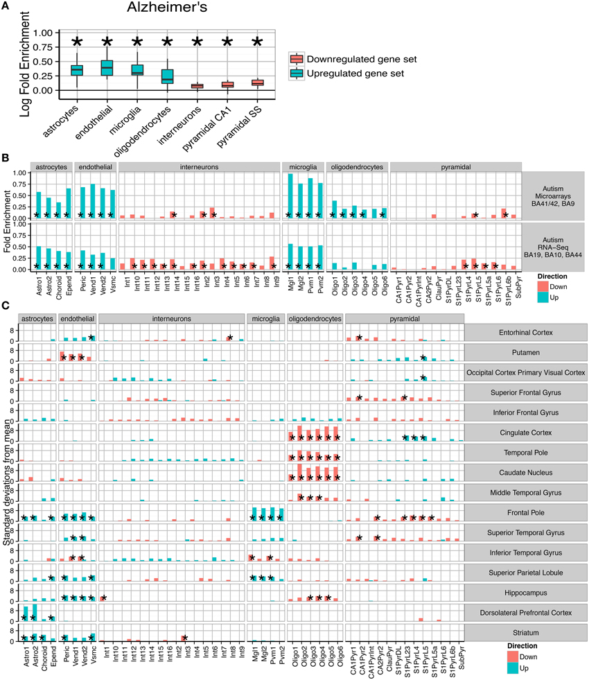
A recent study of gene expression in neurological conditions including autism showed just how broadly disturbed gene expression is.


(A) Consistent fold enrichments were found for each cell type across fourteen cortical and three subcortical brain regions of Alzheimer's patients. The box plots mark the distribution of cellular fold enrichments across all the brain regions examined. Asterisks mark that the fold enrichment for each cell type that was found to be significantly non-zero with p < 0.05. (B) Two independent autism studies show the same cellular phenotypes, including upregulation of glial cells and downregulation of neurons. Asterisks mark those cell types found to be significantly differential with p < 0.05 after BH correction over all groups.


Here I am making the point that even though only a handful of genes may have an identifiable dysfunction, a much broader range of genes seem to be affected, as we see in the wide range of over and under expressed genes.

While it would be logical to think about a specific dysfunction needing a therapy that targets just that gene, this appears not to be necessary.

It appears that downstream processes may be the most damaging/relevant, for example disturbances in Protein Kinase A and C (PKA and PKC) may play a key role in many cases of regressive autism, and this will feature in its own post, because it would be treatable today. 

Reduced activity of protein kinase C in the frontal cortex of subjects with regressive autism: relationship with developmental abnormalities.


Brain Region–Specific Decrease in the Activity and Expression of Protein Kinase A inthe Frontal Cortex of Regressive Autism


Both the above papers are by Abha and Ved Chauhan.  I put Abha on my Dean’s list long ago.  I did have a discussion with her a while back.  She is clearly a very nice person and intellectually towers over the Curemark lady (Joan Fallon) who gets $40 million to play with her pancreatic enzymes, but never publishes anything except very superficial patents.


I think for $40 million Abha and Ved could figure it all out.

PKB, otherwise known as Akt is also very relevant to some types of autism.

Tamoxifen, recently shown to reverse autism in a SHANK3 mouse model, is a PKC inhibitor.

Another epigenetic drug, Theophylline activates PKA.

Akt, also known as protein kinase B (PKB), is a central node in cell signaling downstream of growth factors, cytokines, and other cellular stimuli. Aberrant loss or gain of Akt activation underlies the pathophysiological properties of a variety of complex diseases, including type-2 diabetes and cancer.

If you could identify if a particular person was hypo/hyper in PKA, PKB and PKC, this might well open the door to an effective treatment.


Research on PKB, also known as AKT

Dysregulation of theIGF-I/PI3K/AKT/mTOR signaling pathway in autism spectrum disorders.


And a paper from the clever Japanese:-


Autism spectrum disorder is a set of neurodevelopmental disorders in terms of prevalence, morbidity and impact to the society, which is characterized by intricate behavioral phenotype and deficits in both social and cognitive functions. The molecular pathogenesis of autism spectrum disorder has not been well understood, however, it seems that PI3K, AKT, and its downstream molecules have crucial roles in the molecular pathogenesis of autism spectrum disorder. The PI3K/AKT signaling pathway plays an important role in the regulation of cell proliferation, differentiation, motility, and protein synthesis. Deregulated PI3K/AKT signaling has also been shown to be associated with the autism spectrum disorder. Discovery of molecular biochemical phenotypes would represent a breakthrough in autism research. This study has provided new insight on the mechanism of the disorder and would open up future opportunity for contributions to understand the pathophysiology


For those who favour dietary intervention:-


Based on the above chart curcumin should likely be good for my N=1 case of autism. Time will tell.


Consequences of upstream dysfunctions

So it might be better to consider autism as a disease of wider downstream gene expression, rather than necessarily of “faulty” genes.  Modulating the resulting wider gene expression may be much more realistic than fixing individual genes.

It is certainly plausible that the body has its own protective self-repair mechanism that might be somehow re-energized. Some people have pondered why so many highly intelligent mathematicians and computer scientists seem have relatives with autism.  The clever genes do associate with a type of autism plus ID/MR.  It was suggested that protective genetic changes might be in play, so that the people with the most genetic variance are actually the family members without the autism.

This does remain conjecture, but as more whole genome data is collected we are seeing some interesting findings.

A fascinating very recent study that looked at a group of 53 families with autism using the traditional approach of whole exome sequencing and also microarray. 

Using these methods, that are the current gold standard, the researchers found very little.  Dysfunctions in the 700 known autism genes were not detected.

However using more expensive whole genome sequencing, dysfunctions were identified in the “DNA junk” zone very close beside the known autism genes.  The researchers were then able to identify the genetic cause of 30% of the cases, a big improvement on 0%.  I expect if they looked a little harder the 30% would be higher.


“We performed whole-genome sequencing (WGS) of 208 genomes from 53 families affected by simplex autism.”

“For the majority of these families, no copy-number variant (CNV) or candidate de novo gene-disruptive single-nucleotide variant (SNV) had been detected by microarray or whole-exome sequencing (WES).

Comparing the sequences of the individuals with autism and those of their unaffected siblings, the researchers found that people with autism are more likely to have genetic variants — either single base-pair changes in the sequence or small CNVs — in swaths of DNA abutting known autism genes. But the researchers only found the variants after they restricted their search to regions of the genome already implicated in autism, and even then the statistical significance is modest.

Sequencing whole genomes could reveal the genetic cause of autism in as much as 30 percent of people for whom faster and cheaper sequencing methods come up short

“It’s increasing power even in areas that are supposed to be covered by whole-exome sequencing,” says Peixoto. “It seems that it’s clear that whole-genome sequencing will become the standard.”


One specific microRNA has strong links to autism spectrum disorder, say TSRI scientists


Epigenopathies

Many diseases have an epigenetic component. The severe progressive asthma that is COPD is a well-known example.  It appears that smoking in middle age often leads to permanent epigenetic changes that come back to haunt often then non-smokers in old age.  Even though they have not smoked for twenty years, there oxidative stress response has been permanently modified.  This results in a kind of steroid resistance, so that usually reliable drug therapies fail to work. 

It is thought that autism has an epigenetic component.  This would do some way to explaining 30-40% of the increase in prevalence in recent years that is not explained by ever widening diagnostic criteria.

Because epigenetic changes can be heritable and can be accumulated from all kinds of exposures, even simple ones like severe emotional stress and pollution, you can reconcile autism as being primarily a genetic condition even though incidence has clearly risen within one or two generations. So you can have an “epigenetic epidemic”, so to speak.


Epigenetics as a therapy

While much is written about epigenetic change being bad, it could also be good.

There are many known substances that affect gene expression; some are very target specific which is useful.

This answers a recent issue raised by a reader of this blog who did exome sequencing. What is the point of discovering a genetic dysfunction if there is no therapy? Medicine is some decades behind science, better to know what gene is affected because you well be able to affect its expression, you just need some help from Google.

Epigenetic therapy could be used to remove unwanted tags, but it could also be used to leave new ones to upregulate under-expressed genes.

Such epigenetic therapy is already a reality in COPD and is being considered for rare single autisms where one copy of the gene is not functional, so turn up the volume on the remaining copy.

As we saw in the post on epigenetics, one potential category of drugs are HDAC inhibitors, these would affect one epigenetic mechanism.

There are many such HDAC inhibitors and most have other modes of action, so you cannot be sure what is giving the noted effect.


Valproate

This epilepsy drug has numerous effects including as a HDAC inhibitor.  Given to mothers during pregnancy it can cause autism in the offspring, but when given to the affected offspring the autism can be reduced.

Valproate is given off label to treat autism even when no epilepsy is present.

As we saw in the comments section, long term valproate se can have side effects.


Sulforaphane

This substance derived from broccoli and patented by Johns Hopkins, is another HDAC inhibitor.  It also upregulates Nrf2, which turns on the oxidative response genes.  This was proposed as a COPD therapy by Professor Barnes.

We saw in a post that for Nrf2 to have its full effect there needed to be enough of a protein called DJ-1.  You can increase DJ-1 expression with cinnamon (sodium benzoate).

That was one reason to think that cinnamon would complement Sulforaphane as a therapy for both COPD and some autism.


Sodium Butyrate

Sodium Butyrate is an HDAC inhibitor that is available as a supplement. We came across it in an earlier post as a precursor to butyric acid.  Butyric acid plays a role in the permeability of the gut and the Blood Brain Barrier (BBB).  It also seems to protect from auto immune disease.

Butyrate is fed to millions of farm animals every day to increase their resistance to auto-immune disease.

Butyric acid is produced naturally in the gut by the bacteria living there, however the amount can be increased by the uses of a particular probiotic-bacteria.

This would support the uses of sodium butyrate and the Miyari 588 bacteria.

I have on my to-do-list to investigate higher doses of Miyari 588, but having read the comment by Alli that 500 mg of sodium butyrate is effective, I will try that first.  She also found higher doses ineffective, which was the same in a mouse study published last November,

The study below highlights which genes were down-regulated and which were up-regulated, the overall effect was beneficial


Sodium butyrate attenuate ssocial behavior deficits and modifies the transcription ofinhibitory/excitatory genes in the frontal cortex of an autism model.


The core behavioral symptoms of Autism Spectrum Disorders (ASD) include dysregulation of social communication and the presence of repetitive behaviors. However, there is no pharmacological agent that is currently used to target these core symptoms. Epigenetic dysregulation has been implicated in the etiology of ASD, and may present a pharmacological target. The effect of sodium butyrate, a histone deacetylase inhibitor, on social behavior and repetitive behavior, and the frontal cortex transcriptome, was examined in the BTBR autism mouse model. A 100 mg/kg dose, but not a 1200 mg/kg dose, of sodium butyrate attenuated social deficits in the BTBR mouse model. In addition, both doses decreased marble burying, an indication of repetitive behavior, but had no significant effect on self-grooming. Using RNA-seq, we determined that the 100 mg/kg dose of sodium butyrate induced changes in many behavior-related genes in the prefrontal cortex, and particularly affected genes involved in neuronal excitation or inhibition. The decrease in several excitatory neurotransmitter and neuronal activation marker genes, including cFos Grin2b, and Adra1, together with the increase in inhibitory neurotransmitter genes Drd2 and Gabrg1, suggests that sodium butyrate promotes the transcription of inhibitory pathway transcripts. Finally, DMCM, a GABA reverse agonist, decreased social behaviors in sodium butyrate treated BTBR mice, suggesting that sodium butyrate increases social behaviors through modulation of the excitatory/inhibitory balance. Therefore, transcriptional modulation by sodium butyrate may have beneficial effects on autism related behaviors.


Theophylline

Theophylline is an old asthma drug that is an HDAC inhibitor.

At low doses it is now being trialled as an epigenetic add-on therapy in COPD.  It pretty obviously does work, but data needs to be collected to measure how effective it is and what is the best dose.

It shows how the COPD researchers/clinicians like Professor Barnes are doing a good job and not frightened to experiment.

Would a similar low dose of theophylline benefit a sub-group of those with autism/schizophrenia?  I think it is quite likely.

COPD and autism/schizophrenia share the same impaired oxidative stress response.


Chronic Obstructive Pulmonary Disease (COPD) is a progressive lung disease characterised by progressive airflow limitation. In the UK, it affects around 3 million people, is the fifth leading cause of death and costs the NHS approximately £1 billion annually. Exacerbations of COPD account for 60% of NHS COPD costs and are associated with accelerated rate of lung function decline, reduced physical activity, reduced quality of life, increased mortality and increased risk of co-morbidities. COPD treatment guidelines recommend inhaled corticosteroids (ICS) to reduce exacerbations and improve lung function. However, in COPD, airway inflammation is relatively insensitive to the anti-inflammatory effects of ICS and even high doses fail to prevent exacerbations. Preclinical and pilot studies demonstrate that low dose theophylline may increase the sensitivity of the airway inflammation to ICS, and thus when used with ICS will reduce the rate of COPD exacerbation. In this study we will determine the clinical effectiveness and cost-effectiveness of adding low dose theophylline to ICS therapy in patients with COPD. The primary outcome is the number of exacerbations. The primary economic outcome is the cost-per-QALY gained during the one year treatment period. We will recruit 1424 participants from primary and secondary care across seven areas of the UK. Participants will be randomised to theophylline (200 mg once or twice daily depending on smoking status and weight) or placebo for 12 months. We will follow participants up at six and twelve months to assess the number of exacerbations. We will also collect data on adverse events, health care utilisation, quality of life and breathlessness, and lung function. Low dose theophylline is cheap (10p/day) and, if shown to make current ICS therapy more effective in a cost effective manner, it will improve the quality of life of COPD patients and reduce the burden of COPD on the NHS.


At large doses, Theophylline has long been a therapy for asthma and COPD, but as with Sodium Butyrate, it is quite possible that larger doses of Theophylline produce a different result.  In other words the epigenetic effect fortunately comes from the low dose.

Low doses mean less chance of side effects.

For example, in anyone predisposed to reflux/GERD/GORD many asthma drugs pose a problem because at the same time as opening the airways in your lungs they will relax the lower esophageal sphincter and allow stomach acid to rise upwards.

We saw in an earlier post that in some types of autism something called mGluR5 is dysfunctional in the brain. By chance mGluR5 is also involved in closing the lower esophageal sphincter.  In people with reflux/GERD/GORD a mGluR5 inhibitor was found to have promise for the management of their symptoms.


Randomised clinical trial:effects of monotherapy with ADX10059, a mGluR5 inhibitor, on symptoms and reflux events in patients with gastro-oesophageal reflux disease.


So it is not surprising that many people with autism also have reflux/GERD/GORD. 

But the dysfunction with mGluR5 in autism can be both hyper and hypo, so the therapy might be a positive allosteric modulator (PAM), or a negative allosteric modulator (NAM).  

In someone with autism + reflux/GERD/GORD  it would be reasonable to think a NAM, like ADX10059, might help both conditions.


Gene Repression and Genome Stability

There is another epigenetic process that may be disturbing gene expression in some people and may be treatable.

I have been trying to find why so many people with autism can benefit from biotin; I think I have found a plausible explanation.

“Biotinylation of histones plays a role in gene repression and repression of transposable elements, thereby maintaining genome stability”

I think in some people with autism and no clinical deficiency of biotin the continued “overdosing” of biotin might be having an effect on gene expression, bringing things a little closer to where they should be.

Rather beyond the scope of this blog, it appears that in some people the impaired genome stability, reversible with biotin(ylation), this might be a significant cancer risk.

In essence, for most people supraphysiological concentrations of biotin will do absolutely nothing, but in a sub-group it might do a lot of good.  It is epigenetic, but you do not have to understand it to benefit from it.  It is complicated.


Transposable elements such as long terminal repeats (LTR) constitute 45% of the human genome; transposition events impair genome stability. Fifty-four promoter-active retrotransposons have been identified in humans. Epigenetic mechanisms are important for transcriptional repression of retrotransposons, preventing transposition events, and abnormal regulation of genes. Here, we demonstrate that the covalent binding of the vitamin biotin to lysine-12 in histone H4 (H4K12bio) and lysine-9 in histone H2A (H2AK9bio), mediated by holocarboxylase synthetase (HCS), is an epigenetic mechanism to repress retrotransposon transcription in human and mouse cell lines and in primary cells from a human supplementation study. Abundance of H4K12bio and H2AK9bio at intact retrotransposons and a solitary LTR depended on biotin supply and HCS activity and was inversely linked with the abundance of LTR transcripts. Knockdown of HCS in Drosophila melanogaster enhances retrotransposition in the germline. Importantly, we demonstrated that depletion of H4K12bio and H2AK9bio in biotin-deficient cells correlates with increased production of viral particles and transposition events and ultimately decreases chromosomal stability. Collectively, this study reveals a novel diet-dependent epigenetic mechanism that could affect cancer risk.

Here, we provide evidence for the existence of a novel diet-dependent epigenetic mechanism that represses retrotransposons. Importantly, we demonstrated that depletion of biotinylated histones in biotin-deficient cells increases LTR transcript levels, production of viral particles, and retrotransposition events, and ultimately decreases chromosomal stability. Both biotin deficiency and supplementation are prevalent in the US. For example, moderate biotin deficiency has been observed in up to 50% of pregnant women (35,36). About 20% of the US population reports taking biotin supplements (37), producing supraphysiological concentrations of vitamin in tissues and body fluids (23,28,35). The findings presented here suggest that altered biotin status in these population subgroups might affect chromosomal stability and cancer risk. 

Biotin and biotinidase deficiency


Biotin requirements for DNA damage prevention


Conclusion

I never got round to writing part 2 of my epigenetics post, but my experience of HDAC inhibitors to date has been very positive.

I would be the first to admit that this is rather hit and miss.  It was only when reading the paper on potential therapies for Pitt Hopkins, that was openly musing about HDAC inhibitors, in an equally hit and miss approach, that I thought I would write further about it.

It really seems totally haphazard, because you cannot predict the effect with any level of certainty.  If there is a self-repair mechanism trying to maintain homeostasis of the genome, haphazard may be good enough.

10mg of biotin twice a day does have a mild but noticeable stabilizing effect; is this caused by better maintaining genome stability? I have no idea. 

I will try sodium butyrate and if it works I will have to establish what dose of Miyari 588 produces the same effect.  Both are used in animal feed to reduce inflammatory disease, so you are already indirectly exposed to them if you eat meat.

Theophylline should also be investigated.  This is a very well understood drug and small doses really do seem to help people with COPD.

PKA, PKB and PKC are likely at the core of most people’s autism.  Many existing therapies can modify their expression.

Whole genome sequencing, carried out at great precision, is clearly the only satisfactory genetic testing method.  The other, cheaper, methods are just missing key data and giving many false negative results, i.e. saying there are no identifiable genetic dysfunctions, when this is not true.


Thursday 3 September 2015

Gene Silencers and Enhancers in Autism; plus Epicatechin, MOCOS, Ferritin and Oxidative Stress (GR, GPx, GCL, GCLM)


The original idea of this blog was to try to keep complicated things as simple as possible, so at times things may get over-simplified.  

This post starts out simple and then gets rather involved in oxidative stress.

When people think about genes, they are nearly always thinking about the “blueprints” that are encoded on your DNA.  As it turns out only about 5% of your DNA is dedicated to this function; this 5% is contained in the exome.

Much autism research is dedicated to finding faulty “blueprints” that might account for autism.  There are now several hundred so called “autism genes”, where an error in the “blueprints”, means that the associated protein is not produced to its intended specification.

We also have seen that genetic defects just lead to a possibility of something going wrong.  A “faulty gene” creates the possibility of a specific dysfunction happening, it does not mean 100% that it will happen. 


Partial dysfunctions and partial deficiency

We also saw that even when a single gene dysfunction, like for fragile-X, occurs it does not always cause a catastrophic failure, rather it produces a spectrum from mild to severe.

This point is important since it seems in autism there can often be “partial dysfunctions” leading to “partial deficiencies”.  This is just a less severe form of the “rare” total dysfunctions.  The growing list of examples includes partial biotinidase deficiency, partial glutathione reductase deficiency and partial glutathione peroxidase deficiency.  Today we will also encounter ferritin (iron storage) partial deficiencies.  In a future post we will look the vitamin B12 partial dysfunction that occurs in about a quarter of schizophrenia and autism cases.

This then leads us to the subject of gene expression, which means how much, where, when and how often a gene is turned “on”.  This is actually what really matters, since even perfectly good genes, when over-expressed, can do great damage.  We saw that in the case of Down Syndrome there is about 50% over expression in up to 300 genes.  In the case of Down Syndrome the reason for this overexpression lies in the exome.  In effect there is a double set of blueprints for those 300 genes.

Within the remaining 95% of your DNA are so-called enhancers and silencers.  Their job is to determine which genes are turned on (enhancers) or turned off (silencers) in which part of the body.  So a gene might encode a calcium channel, but that calcium channel should only be in certain parts of the body and only to a certain degree.  We need the correct clean blueprint and we need it applied in the right part of the body and only to the desired extent.

I was very pleased to see that some scientists have started to look at the role of enhancers in autism.  I have already noticed that some substances that are known to affect gene expression are particularly effective in autism.  This suggests to me that in some types of autism, the problem may actually be simply in gene expression rather than any faulty genetic “blueprint”.

Now the science of enhancers and particularly silencers is still at the emerging stage, but the research showed that in at least 100 locations, there were significant anomalies in those with autism.


This is an easy to read summary of the research paper below.


Abstract

Despite major progress in identifying enhancer regions on a genome-wide scale, the majority of available data are limited to model organisms and human transformed cell lines. We have identified a robust set of enhancer RNAs (eRNAs) expressed in the human brain and constructed networks assessing eRNA-gene coexpression interactions across human fetal brain and multiple adult brain regions. Our data identify brain region-specific eRNAs and show that enhancer regions expressing eRNAs are enriched for genetic variants associated with autism spectrum disorders.


We also have the removable markers on the 5% of DNA that cause epigenetic changes.  This is another way of turning on or off specific genes.  These markers can be caused by environment factors like smoking, or even stress, these markers are potentially both removable and inheritable.     

The emerging science of Proteomics is the study of gene expression itself, so it is measuring all the proteins that the genes actually produced.


Limits of Genetic Testing

So while in some cases genetic testing of the 5% of DNA usually examined may indeed be useful, if your problem was in the other 95% of DNA it will not help.

To be useful in autism you would need to measure gene expression in the brain or the local activity of the enhancers/silencers, since it varies throughout the body.  In the Australian study above they measured the enhancer activity in the brain, by looking for the special enhancer molecules the enhancers produce.

This is all way beyond the scope of this blog.

However when I see “safe” substances like Sulforaphane, Epicatechin and even statins that are known to affect the expression of multiple genes, I take note. 

Steroids also affect gene expression, but great care has to be taken with steroids.

Statins have numerous interesting effects in the brain and in cancer cells.  In autism they have an effect on PTEN and BCL2 for example.


The observed impact of pravastatin on gene expression may explain the pleiotropic effects of statins when they are used as adjuvants in chemotherapy and suggests impact on gene expression as a possible cause of side effects from statin use.


As pointed out in the last paper, changing gene expression can be bad as well as good.  It all depends where you are starting from and what genes you want to enhance/silence.


Other therapies to modify gene expression

Today’s scientific knowledge does not always allow us to target the expression of specific genes, this very much remains future science.

However, the remarkable effects of some substances, in some people, does suggest some options.  As is often the case this takes us back to oxidative stress, which does seem to affect many conditions and is quite well studied. There is no shortage of anecdotal evidence.

We know from the research that oxidative stress is ever-present in autism and that people with autism are particularly sensitive to it.

One substance previously mentioned in this blog, epicatechin, is known to change the expression of many genes including STAT1, MAPKK1, MRP1, and FTH1, which are involved in the cellular response to oxidative stress.


Ferritin

Rather off subject the FTH1 gene encodes the heavy subunit of ferritin, the major intracellular iron storage protein.


Children with autism spectrum disorders had significantly lower ferritin levels compared with controls
Within the autism spectrum disorders population, median ferritin levels were significantly lower in patients with poor sleep efficiency (7 ng/mL) versus those with normal sleep efficiency (29 ng/mL) (P = 0.01).


Low ferritin would indicate an iron storage problem and likely anemia/anaemia

Low ferritin has many effects, including surprisingly, poor sleeping patterns.
  
Is it such a surprise that a cup of cocoa (epicatechin) before bed used to be given to ensure a good night’s sleep?  (all via FTH1, I presume)

Perhaps poor sleep in autism is just another consequence of oxidative stress?


MOCOS

In the recent paper on MOCOS:-


I noted that:-

Furthermore, we found that MOCOS misexpression induces increased oxidative-stress sensitivity.

MOlybdenum COfactor Sulfurase (MOCOS), is an enzyme involved in purine metabolism and a newly identified player in ASD. MOCOS appears to be downregulated in autism and this has multiple effects, one being increased sensitivity to oxidative stress.


Seemingly unknown to the French MOCOS researchers, there already is a therapy:-


Since I do not have any of the above biosynthetic precursor at hand, but I do have high flavanol cocoa in the kitchen, it is time to look again at epicatechin.


Epicatechin

There are two very similar substances catechin and epicatechin; both are flavonoids.  Both affect gene expression and both seem to have numerous good properties.

Epicatechin is found in large quantities in mildly processed cocoa, which catechin in found in large quantities in certain types of Chinese tea.

We saw in an earlier post that Mars, the chocolate company, has invested substantially in the science of cocoa and its flavonoids.  They have just signed a 5 year research contract with Harvard.

Catechin affects the fat metabolism and is therefore a potential therapy for obesity.  Oolong tea has been shown to have this effect, but you do need to drink a great deal of it.


CONCLUSIONS:
Oolong tea could decrease body fat content and reduce body weight through improving lipid metabolism. Chronic consumption of oolong tea may prevent against obesity.


ABSTRACT Various health benefits of the cocoa flavanol (-)-epicatechin (EC) have been attributed to its antioxidant and anti-inflammatory potency. In the present study we investigated whether EC is able to prevent deterioration of the anti-inflammatory effect of the glucocorticoid (GC) cortisol in the presence of oxidative stress. It was found that cortisol reduces inflammation in differentiated monocytes. Oxidative stress extinguishes the anti-inflammatory effect of cortisol, leading to cortisol resistance. EC reduces intracellular oxidative stress as well as the development of cortisol resistance. This further deciphers the enigmatic mechanism of EC by which it exerts its anti-inflammatory and antioxidant action. The observed effect of the cocoa flavanol EC will especially be of relevance in pathophysiological conditions with increased oxidative stress and consequential GC resistance and provides a fundament for the rational use of dietary antioxidants


Abstract
Background: Consumption of flavonoid-rich beverages, including tea and red wine, has been associated with a reduction in coronary events, but the physiological mechanism remains obscure. Cocoa can contain extraordinary concentrations of flavanols, a flavonoid subclass shown to activate nitric oxide synthase in vitro.
Objective: To test the hypothesis that flavanol-rich cocoa induces nitric-oxide-dependent vasodilation in humans.
Design: The study prospectively assessed the effects of Flavanol-rich cocoa, using both time and beverage controls. Participants were blinded to intervention; the endpoint was objective and blinded.
Methods: Pulse wave amplitude was measured on the finger in 27 healthy people with a volume-sensitive validated calibrated plethysmograph, before and after 5 days of consumption of Flavanol-rich cocoa [821 mg of flavanols/day, quantitated as (−)-epicatechin, (+)-catechin, and related procyanidin oligomers]. The specific nitric oxide synthase inhibitor, NG-nitro-l-arginine methyl ester (l-NAME) was infused intravenously on day 1, before cocoa, and on day 5, after an acute ingestion of cocoa.
Results: Four days of flavanol-rich cocoa induced consistent and striking peripheral vasodilation (P = 0.009). On day 5, pulse wave amplitude exhibited a large additional acute response to cocoa (P = 0.01). l-NAME completely reversed this vasodilation (P = 0.004). In addition, intake of flavanol-rich cocoa augmented the vasodilator response to ischemia. Flavanol-poor cocoa induced much smaller responses (P = 0.005), and none was induced in the time-control study. Flavanol-rich cocoa also amplified the systemic pressor effects of l-NAME (P = 0.005).
Conclusion: In healthy humans, flavanol-rich cocoa induced vasodilation via activation of the nitric oxide system, providing a plausible mechanism for the protection that flavanol-rich foods induce against coronary events.


Abstract

The Kuna Indians, who reside in an archipelago on the Caribbean Coast of Panama, have very low blood pressure (BP) levels, live longer than other Panamanians, and have a reduced frequency of myocardial infarction, stroke, diabetes mellitus, and cancer—at least on their death certificates. One outstanding feature of their diet includes a very high intake of flavanol-rich cocoa. Flavonoids in cocoa activate nitric oxide synthesis in healthy humans. The possibility that the high flavanol intake protects the Kuna against high BP, ischemic heart disease, stroke, diabetes mellitus, and cancer is sufficiently intriguing and sufficiently important that large, randomized controlled clinical trials should be pursued.


Glutathione reductase (GR) and (partial) Glutathione reductase deficiency

Glutathione reductase (GR) catalyzes the reduction of glutathione disulfide (GSSG) to the sulfhydryl form glutathione (GSH), which is a critical molecule in resisting oxidative stress and maintaining the reducing environment of the cell.

Glutathione reductase reduces one mole of GSSG to two moles of GSH.

Glutathione reductase deficiency is a “rare” disorder in which the glutathione reductase activity is absent from erythrocytes, leukocytes or both. In one study this disorder was observed in only two cases in 15,000 tests for glutathione reductase deficiency performed over the course of 30 years. In the same study, glutathione reductase deficiency was associated with cataracts and favism in one patient and their family, and with severe unconjugated hyperbilirubinemia in another patient. It has been proposed that the glutathione redox system (of which glutathione reductase is apart) is almost exclusively responsible for the protecting of eye lens cells from hydrogen peroxide because these cells are deficient in catalase, the enzyme which catalyzes the breakdown of hydrogen peroxide, and the high rate of cataract incidence in glutathione reductase deficient individuals.

Some patients exhibit deficient levels of glutathione activity as a result of not consuming enough riboflavin in their diets. Riboflavin is a precursor for FAD, whose reduced form donates two electron to the disulfide bond which is present in the oxidized form of glutathione reductase in order to begin the enzyme's catalytic cycle.
In 1999, a study found that 17.8% of males and 22.4% of females examined in Saudi Arabia suffered from low glutathione reductase activity due to riboflavin deficiency.


Abstract

Glutathione reductase (GR) is a ubiquitous enzyme required for the conversion of oxidized glutathione (GSSG) to reduced glutathione (GSH) concomitantly oxidizing reduced nicotinamide adenine dinucleotide phosphate (NADPH) in a reaction essential for the stability and integrity of red cells. Mutations in the GR gene and nutritional deficiency of riboflavin, a co-factor required for the normal functioning of GR, can cause GR deficiency. We conducted a study on 1691 Saudi individuals to determine the overall frequency of GR deficiency and to identify whether the deficiency results from genetic or acquired causes or both. The activity of GR was measured in freshly prepared red cell haemolysate in the presence and absence of flavin adenine dinucleotide (FAD) and the activity coefficient (AC) was determined. Samples with low GR activity (> 2.0 IU/g haemoglobin) both in the presence and absence of FAD and an AC between 0.9 and 1.2 were considered GR-deficient. Samples with AC > or = 1.3 were considered riboflavin-deficient. The overall frequency of partial GR deficiency was 24.5% and 20.3% in males and females respectively. In addition, 17.8% of males and 22.4% of females suffered from GR deficiency due to riboflavin deficiency. This could be easily corrected by dietary supplementation with riboflavin. No cases of severe GR deficiency were identified.


Regular readers may recall something very similar with biotin and its enzyme biotinidase.  Biotinidase deficiency is supposedly such a rare metabolic disorder that it is no longer screened for; however, in an autism study in Crete, Greece it was found that partial biotinidase deficiency was quite common.


Glutathione peroxidase

Glutathione peroxidase (GPx) is the general name of an enzyme family with peroxidase activity whose main biological role is to protect the organism from oxidative damage.
The biochemical function of glutathione peroxidase is to reduce lipid hydroperoxides to their corresponding alcohols and to reduce free hydrogen peroxide to water.

In earlier posts on anti-oxidants we saw the following presentation from the German scientist.  Note Glutathione (GSH) peroxidases, left halfway down


Glutamate Cysteine Ligase (GCL)

  
Glutamate Cysteine Ligase (GCL) is the first enzyme of the cellular glutathione (GSH) biosynthetic pathway.

GSH, and by extension GCL, is critical to cell survival.

Nearly every eukaryotic cell, from plants to yeast to humans, expresses a form of the GCL protein for the purpose of synthesizing GSH

Dysregulation of GCL enzymatic function and activity is known to be involved in the vast majority of human diseases, such as diabetes, Parkinson's disease, Alzheimers disease, COPD, HIV/AIDS, and cancer. This typically involves impaired function leading to decreased GSH biosynthesis, reduced cellular antioxidant capacity, and the induction of oxidative stress.


Measuring GR, GPx, GCL in Autism

Fortunately somebody has already measured GR, GPx and GCL in autism, and not surprisingly they are all dysfunctional.  The paper is by the Chauhans, who already feature on my Dean’s list of researchers.


In the cerebellum tissues from autism (n=10) and age-matched control subjects (n=10), the activities of GSH-related enzymes glutathione peroxidase (GPx), glutathione-S-transferase (GST), glutathione reductase (GR), and glutamate cysteine ligase (GCL) involved in antioxidant defense, detoxification, GSH regeneration, and synthesis, respectively, were analyzed. GCL is a rate-limiting enzyme for GSH synthesis, and the relationship between its activity and the protein expression of its catalytic subunit GCLC and its modulatory subunit GCLM was also compared between the autistic and the control groups. Results showed that the activities of GPx and GST were significantly decreased in autism compared to that of the control group (P<0.05). Although there was no significant difference in GR activity between autism and control groups, 40% of autistic subjects showed lower GR activity than 95% confidence interval (CI) of the control group. GCL activity was also significantly reduced by 38.7% in the autistic group compared to the control group (P=0.023), and 8 of 10 autistic subjects had values below 95% CI of the control group. The ratio of protein levels of GCLC to GCLM in the autism group was significantly higher than that of the control group (P=0.022), and GCLM protein levels were reduced by 37.3% in the autistic group compared to the control group. A positive strong correlation was observed between GCL activity and protein levels of GCLM (r=0.887) and GCLC (r=0.799) subunits in control subjects but not in autistic subjects, suggesting that regulation of GCL activity is affected in autism. These results suggest that enzymes involved in GSH homeostasis have impaired activities in the cerebellum in autism, and lower GCL activity in autism may be related to decreased protein expression of GCLM.

GCLM referred to above is Glutamate-cysteine ligase, it is the first rate limiting enzyme of glutathione synthesis, it is encoded by the GCLM gene. This is an enzyme/ gene you would want to upregulate.
https://en.wikipedia.org/wiki/GCLM

Fortunately we can upregulate GPx enzyme activity with catechin or epicatechin.


Abstract

OBJECTIVES:

The objective of this study was to investigate the effects of catechin and epicatechin on the activity of the endogenous antioxidant enzymes superoxide dismutase (SOD) and glutathione peroxidase (GPx) (as well as the total antioxidant capacity (TAC)) of rats after intra-peritoneal (i.p.) administration.

METHODS:

Twenty-four Wistar rats were randomly divided into two groups: the experimental group which was administered daily with a 1:1 mixture of epicatechin and catechin at a concentration of 23 mg/kg body weight for 10 days and the control group which was injected daily with an equal amount of saline. Blood and urine samples were collected before and after the administration period, as well as 10 days after (follow-up).

RESULTS:

Intra-peritoneal administration of catechins led to a potent decrease in GPx levels and a significant increase in SOD levels. TAC was significantly increased in plasma and urine. Malonaldehyde levels in urine remained stable. In the animals treated with catechins, SOD activity showed a moderate negative correlation with GPx activity.

DISCUSSION:

Boosting the activity of the antioxidant enzymes could be a potential adjuvant approach for the treatment of the oxidative stress-related diseases.


The objective of this study was to determine whether i.p. administration of catechin and epicatechin could affect the activity of the antioxidant enzymes, SOD and GPx, as well as the TAC in RBCs, blood plasma, and urine.
The antioxidant enzymes are agents that promote reactions for the removal of reactive species (e.g. O2,.H2O2, etc.). They constitute the first line of
defense against oxidative stress. In conditions of increased oxidative stress, the upregulation of the enzyme activity or even, a possible protection of the enzymessubstrate could be of great importance.

Oxidative stress disturbing homeostasis can be resolved by the application of catechins and epigallocatechin gallate (EGCG)18 and there is growing evidence that, the protection, offered by flavonoids and their in vivo metabolites, is not mediated primarily by H-donating antioxidant processes, but is likely to be partly mediated through specific actions, within signaling pathways.

Catechin and epicatechin administration modulated the activity of SOD and GPx but the overall TAC of the RBCs and of the rats plasma remained stable.
Catechins are considered as potent antioxidants and many of their biological actions have been attributed to that. It would have been expected that since catechins are potent antioxidants in vitro, they would have exerted their classical hydrogen-donating antioxidant activity leading to an increase in TAC; as it is seen in the TAC of plasma. The modulation of the enzymes activity may provide evidence that, catechins exert their primary antioxidant activity by specific action within specific molecular pathways, rather than as scavengers of free radicals.

Oxidative stress is a prominent feature of many acute and chronic diseases and even of the normal aging process. The normal function of the antioxidant enzymes guarantees the preservation of cell integrity and thus they can be considered as potential therapeutic targets of oxidative stress-related diseases.
Various antioxidants are available for therapeutic use but most of them have failed in clinical studies of diseases correlated with oxidative stress. Our results suggest that catechins exert their activity not only by H-donating antioxidant processes but likely through mechanisms and pathways that directly or indirectly regulate the expression of the enzymatic antioxidants.

The understanding of these pathways could be important, in developing pharmacological strategies against oxidative stress-related diseases.


For those with autism plus GI issues / ulcerative colitis :- 


Abstract
Background. This study was pathway of (−)-epicatechin (EC) in the prevention and treatment of intestine inflammation in acute and chronic rat models. Methods. Intestine inflammation was induced in rats using TNBS. The morphological, inflammatory, immunohistochemical, and immunoblotting characteristics of colon samples were examined. The effects of EC were evaluated in an acute model at doses of 5, 10, 25, and 50 mg/kg by gavage for 5 days. The chronic colitis model was induced 1st day, and treated for 21 days. For the colitis relapse model, the induction was repeated on 14th. Results. EC10 and EC50 effectively reduced the lesion size, as assessed macroscopically; and confirmed by microscopy for EC10. The glutathione levels were higher in EC10 group but decreased COX-2 expression and increased cell proliferation (PC) were observed, indicating an anti-inflammatory activity and a proliferation-stimulating effect. In the chronic colitis model, EC10 showed lower macroscopic and microscopic lesion scores and increase in glutathione levels. As in the acute model, a decrease in COX-2 expression and an increase in PC in EC10, the chronic model this increase maybe by the pathway EGF expression. Conclusion. These results confirm the activity of EC as an antioxidant that reduces of the lesion and that has the potential to stimulate tissue healing, indicating useful for preventing and treating intestine inflammation.


Abstract

We studied a polyphenol-enriched cocoa extract (PCE) with epicatechin, procyanidin B2, catechin, and procyanidin B1 as the major phenolics for its anti-inflammatory properties against dextran sulfate sodium (DSS)-induced ulcerative colitis (UC) in mice. PCE reduced colon damage, with significant reductions in both the extent and the severity of the inflammation as well as in crypt damage and leukocyte infiltration in the mucosa. Analysis ex vivo showed clear decreases in the production of nitric oxide, cyclooxygenase-2, pSTAT-3, and pSTAT1α, with NF-κB p65 production being slightly reduced. Moreover, NF-κB activation was reduced in RAW 264.7 cells in vitro. In conclusion, the inhibitory effect of PCE on acute UC induced by DSS in mice was attenuated by oral administration of PCE obtained from cocoa. This effect is principally due to the inhibition of transcription factors STAT1 and STAT3 in intestinal cells, with NF-κB inhibition also being implicated.


 Here is an excellent paper on oxidative stress.  It is about COPD, but applicable to any condition in which oxidative stress is present.


The following paper would suggest that people with COPD would benefit from epicatechin.

The cocoa flavanol (-)-epicatechin protects the cortisol response.


Abstract

Various health benefits of the cocoa flavanol (-)-epicatechin (EC) have been attributed to its antioxidant and anti-inflammatory potency. In the present study we investigated whether EC is able to prevent deterioration of the anti-inflammatory effect of the glucocorticoid (GC) cortisol in the presence of oxidative stress. It was found that cortisol reduces inflammation in differentiated monocytes. Oxidative stress extinguishes the anti-inflammatory effect of cortisol, leading to cortisol resistance. EC reduces intracellular oxidative stress as well as the development of cortisol resistance. This further deciphers the enigmatic mechanism of EC by which it exerts its anti-inflammatory and antioxidant action. The observed effect of the cocoa flavanol EC will especially be of relevance in pathophysiological conditions with increased oxidative stress and consequential GC resistance and provides a fundament for the rational use of dietary antioxidants.


Conclusion

It would seem that in someone with autism epicatechin is worth a try, other indicators might well include:-

·        Low MOCOS
·        Low ferritin
·        Oxidative stress

And even

·        Restless leg syndrome (symptom of low ferritin)
·        Poor sleep patterns (symptom of low ferritin)


Boosting anti-oxidant enzymes (via gene expression) may be a useful add-on therapy to anti-oxidants themselves.  This is likely true for COPD and autism/schizophrenia.

If you are wondering whether there is anemia or iron deficiency in autism, your questions are likely answered here:-


This research considers the prevalence of iron deficiency in children with autism and Asperger syndrome and examines whether this will influence guidelines and treatment. Retrospective analysis of the full blood count and, as far as available, serum ferritin measurements of 96 children (52 with autism and 44 with Asperger syndrome) was undertaken. Six of the autistic group were shown to have iron deficiency anaemia and, of the 23 autistic children who had serum ferritin measured, 12 were iron deficient. Only two of the Asperger group had iron deficiency anaemia and, of the 22 children who had their serum ferritin measured, only three were iron deficient. Iron deficiency, with or without anaemia, can impair cognition and affect and is associated with developmental slowing in infants and mood changes and poor concentration in children. This study showed a very high prevalence of iron deficiency in children with autism, which could potentially compromise further their communication and behavioural impairments.


As we saw with biotin and soon will with vitamin B12, it seems that people with autism can have unexpected deficiencies of key substances even though their diet may not be deficient.  The identified iron deficiency is an iron storage deficiency.  With biotin the body was unable to recycle the vitamin biotin, due to a problem with the enzyme biotinidase, hence there was a deficiency.

Correcting these deficiencies is quite simple and may well improve any related autism symptoms.  In people without these dysfunctions/deficiencies any such supplements would yield no benefit and might even produce side effects.